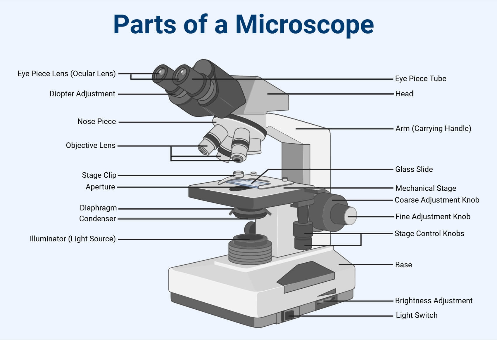
Are you curious about the inner workings of a microscope? Whether you’re a science enthusiast or a student, understanding the basic components of a microscope can be fascinating. Let’s take a closer look at a diagram of a microscope labeled.
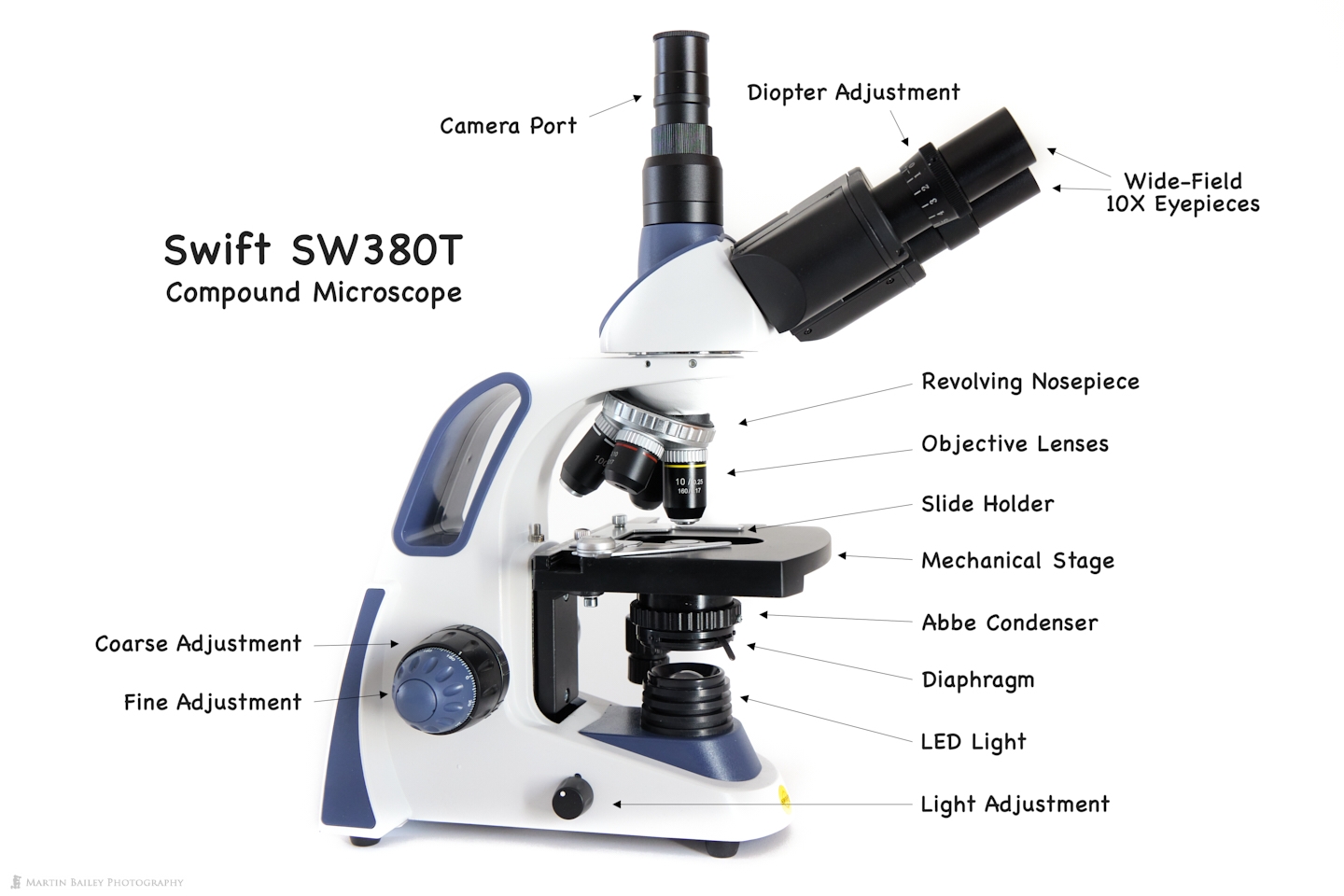
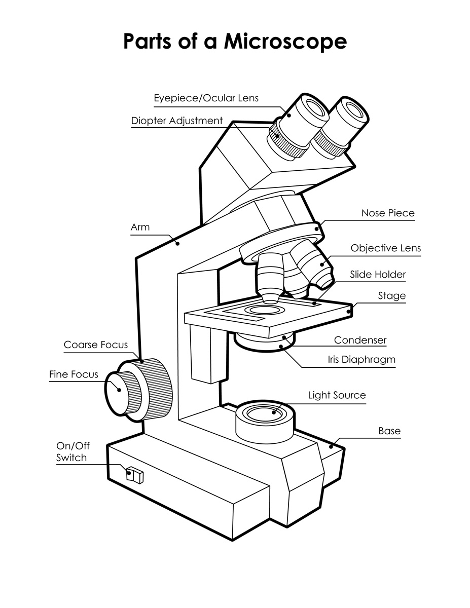
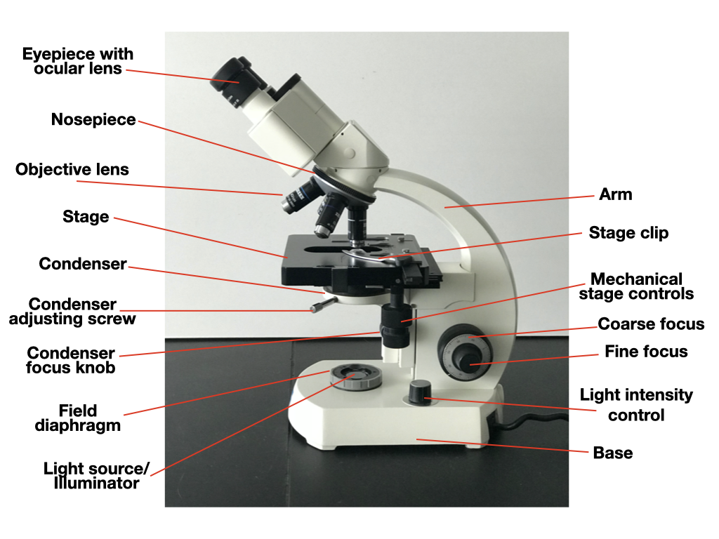
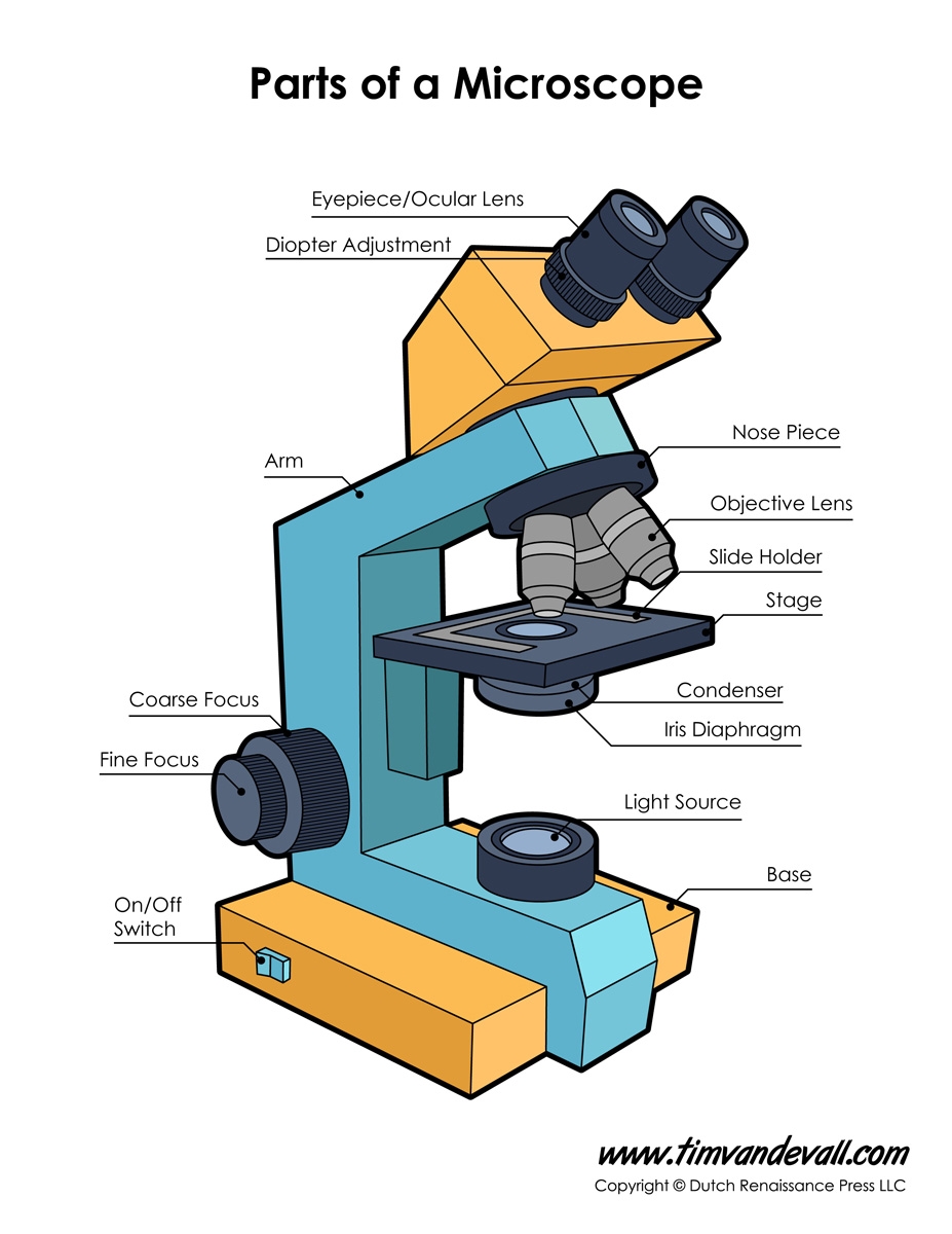
Microscopes are essential tools for scientists, allowing them to observe objects that are too small to be seen with the naked eye. A labeled diagram of a microscope can help you understand how each part contributes to magnifying and illuminating the specimen.
Diagram Of A Microscope Labeled
Diagram Of A Microscope Labeled
At the base of the microscope is the sturdy foot, providing stability during use. The body tube holds the optical components and allows for precise focusing. The nosepiece holds the objective lenses, which can be rotated to change magnification.
The stage is where the specimen is placed for observation, and it may have clips to hold the slide in place. The diaphragm controls the amount of light passing through the specimen, while the light source illuminates the sample from below. The eyepiece, or ocular lens, is where you look through to see the magnified image.
Understanding the various parts of a microscope can enhance your microscopy experience, whether you’re examining cells, bacteria, or other tiny structures. By familiarizing yourself with a labeled diagram of a microscope, you’ll be better equipped to use this powerful tool in your scientific endeavors.
Next time you peer through the lens of a microscope, take a moment to appreciate the intricate design and functionality of this essential scientific instrument. A labeled diagram of a microscope can serve as a helpful guide as you explore the microscopic world around you. Happy exploring!
Microscope Diagram Labeled Unlabeled And Blank Parts Of A Microscope
Microscopy Background College Biology II Laboratory
Microscope Diagram Labeled Unlabeled And Blank Parts Of A Microscope
Parts Of A Microscope With Functions And Labeled Diagram
